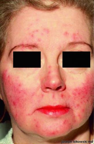
vivid erythema and papules. Pustules are also present, but faint

Frontal view of centrofacial erythema.

Close-up view of centrofacial erythema with scaling

Close-up view of telangiectasias on lateral chin.

Close-up view of papulopustular rosacea.

Phymatous changes on tip of nose, inflammatory lesions (papules and pustules) visible laterally, and telangiectasia.
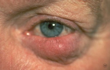
Ocular rosacea

This photo shows bilateral conjunctival redness with eyelid redness and swelling in a patient with ocular rosacea.
bufferLOL
Clincal
Presentation
Symptoms
Physical Exam
Diagnosis and Investigations
Differential
Management
Epidemiology
Pathophysiology
Summary